郭国娜聚焦离子束和扫描电镜的区别是什么意思啊英文
- 聚焦离子束
- 2024-03-25 23:12:24
- 634
fib芯片提供维修、系统安装、技术升级换代、系统耗材,以及应用开发和培训。
Title: Focusing Ion Beam and Scanning Electron Microscopy: A Comparative Study

Introduction
Ion beam and scanning electron microscopy (SEM) are two widely used analytical techniques in materials science and engineering. They belong to the class of microscopy, which is a set of techniques that enable researchers to observe and manipulate objects at a very small scale. In this article, we will discuss the differences between ion beam and scanning electron microscopy and their respective applications in material research.
Ion Beam Microscopy
Ion beam microscopy, also known as ion beam analysis, is a technique that involves the acceleration of ions through a high-energy beam to create a focused beam of ions. The ions are typically generated from various materials, such as metal samples or solutions. The focused beam of ions is then directed onto the sample, where interactions occur between the ions and the material's electrons. These interactions can lead to various chemical changes in the material, resulting in changes in the microstructure, properties, or composition.
One of the main advantages of ion beam microscopy is its ability to analyze a wide range of materials, including metals, semiconductors, and insulators. It is also a highly sensitive technique, capable of detecting changes in the material's microstructure at the atomic level. However, ion beam microscopy also has some limitations. It is time-consuming and requires a high-quality ion beam, which can be challenging to achieve. Additionally, the focused beam can cause damage to the sample, which may affect the results.
Scanning Electron Microscopy
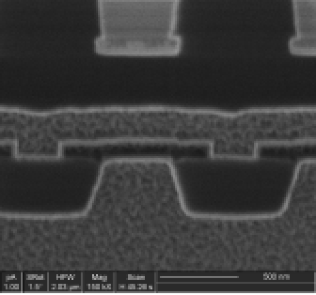
Scanning electron microscopy, on the other hand, is a non-invasive microscopy technique that involves the scanning of a beam of electrons onto the sample. The beam of electrons is directed by a sample stage, which moves slowly across the sample surface. The electrons in the beam collide with the atoms in the sample, causing various effects, such as ionization or chemical changes. These effects enable the observation of the material's microstructure, surface properties, or composition.
SEM has several advantages over ion beam microscopy. It is a faster and more gentle technique, as it does not require the focused beam of ions. It is also a widely used technique for analyzing a wide range of materials, including semiconductors, metals, and insulators. However, SEM also has some limitations, such as its limited depth of analysis and the need for high-quality samples.
Comparison between Ion Beam and Scanning Electron Microscopy
In summary, ion beam and scanning electron microscopy are two powerful analytical techniques that can be used to study the microstructure, properties, and composition of materials. While ion beam microscopy is more sensitive and can analyze a wider range of materials, SEM is faster and more widely used. Both techniques have their respective advantages and limitations, and the choice of technique depends on the specific research question and available resources.
Conclusion
In conclusion, ion beam and scanning electron microscopy are two important analytical techniques that enable researchers to study the microstructure, properties, and composition of materials at a very small scale. While ion beam microscopy is more sensitive and can analyze a wider range of materials, SEM is faster and more widely used. Both techniques have their respective advantages and limitations, and the choice of technique depends on the specific research question and available resources. It is important to note that both techniques can provide valuable insights into the material's properties and composition, which can lead to a better understanding of the material's behavior and performance in different applications.
专业提供fib微纳加工、二开、维修、全国可上门提供测试服务,成功率高!
郭国娜聚焦离子束和扫描电镜的区别是什么意思啊英文 由纳瑞科技聚焦离子束栏目发布,感谢您对纳瑞科技的认可,以及对我们原创作品以及文章的青睐,非常欢迎各位朋友分享到个人网站或者朋友圈,但转载请说明文章出处“聚焦离子束和扫描电镜的区别是什么意思啊英文 ”

上一篇
冷冻扫描电子显微镜多少钱
下一篇
重离子肿瘤治疗优势